Live Cell Imaging for Everyone Using the OpenFlexure Microscope
1 Jun, 2026
To truly understand how a cell heals, moves, or reacts to a new drug, you can’t just take a snapshot; you have to watch it happen live. But live-cell imaging has traditionally come with high barriers to entry. Commercial systems are often prohibitively expensive and represent a large investment for any lab, making is hard or impossible for researchers in low- to middle- income countries (LMICs) to adopt these technologies.
Enter Peter O’Toole, Jodie Malcolm, William Brackenbury, Laura Wiggins, and their network of collaborators across Nigeria, South Africa, and Brazil. They are using an adapted OpenFlexure Microscope (LCI-OFM) to democratise live-cell imaging. By placing these low-cost, 3D-printed microscopes directly into 37°C, humid “oven-style” incubators, the team can continuously monitor dynamic behaviours, such as successfully capturing a 48-hour timelapse of breast cancer cells responding to the chemotherapeutic drug docetaxel.
“Seeing our first 48-hour timelapse come through was bloomin’ amazing!”, Jodie recalls. “After all the work that went into getting the (already brilliant) OpenFlexure microscope running live-cell imaging experiments reliably inside a tissue culture incubator, it felt like a real team victory. Being able to then quantify the data collected on this 3D-printed microscope in a meaningful way using our established CellPhe toolkit pipeline was a real moment of joy and excitement.”
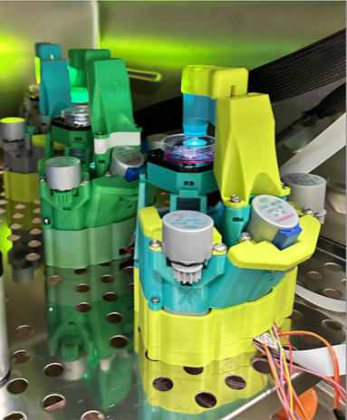
However, making a 3D-printed plastic microscope survive inside a hot, humid incubator required brilliant, community-driven engineering. To withstand the environment, the team relocated sensitive electronics outside the chamber, modified the motor control to reduce heat, and switched to heat-resistant ASA plastic to prevent focus drift.
The team chose the OpenFlexure Microscope precisely because it strikes a rare balance: low cost, high modularity, and complete freedom to customise every part of the system. Its compact footprint was essential for fitting onto an incubator shelf, and its open-design ethos meant it could be adapted exactly to their experimental needs
A Resilient System
The resulting instrument isn’t just affordable; it is built for real-world resilience. The LCI-OFM can run completely offline via a custom graphical user interface, and can even operate seamlessly off a standard power bank, ensuring that crucial experiments aren’t lost during power grid blackouts.
Reliable power is a real challenge for many of the team’s colleagues in Nigeria, South Africa, and Brazil, where planned and unplanned brownouts and blackouts are a routine part of laboratory life. While collaborators have not yet had to rely on the power bank during active experiments, testing in York demonstrated that the system can bridge outages without losing long-term imaging data, a level of resilience that is absolutely essential for researchers working in resource-constrained settings.

Building a Global Network Through Open Science
For the global community, this changes everything. In high-stakes research environments, gaining access to major live-cell imaging centres requires “proof of concept” data - data that researchers can now generate on their own. By lowering the financial and technical barriers, the OpenFlexure community isn’t just serving an existing market; they are helping to create a new one.
The LCI-OFM collaboration grew organically through the open-science community, with connections forming through international meetings, imaging networks, and longstanding partnerships. Training exchanges, including a visit to York supported by the Wellcome Trust and Global BioImaging, helped build advanced microscopy capacity. These relationships continue to strengthen imaging communities across Africa and South America.
South Africa: High-Containment, Lower Risk
The team are now working with Mandy Mason’s group in South Africa to eventually bring the OpenFlexure Microscope into BSL-3 (Cat 3) laboratories.
In these high-containment environments, equipment can be permanently lost if contamination occurs. Having a microscope that is inexpensive, easy to sterilise, and straightforward to replace is a major advantage. Because the system is incubator-safe and humidity-tolerant, most sensitive components remain protected outside the chamber. In a worst-case scenario, only the 3D-printed chassis and motors would need to be swapped out, dramatically reducing both financial risk and workflow disruption. While the system has not yet been deployed in active BSL-3 work, its potential is already reshaping how imaging workflows are planned in these biohazard environments.

Brazil: Watching Infection Happen in Real Time
In Rio de Janeiro, Ana Paula Lima’s team studies Leishmania, the parasites responsible for leishmaniasis, a disease that can take hold when these tiny organisms slip inside our immune cells. The group focuses on how Leishmania promastigotes invade macrophages, the very cells meant to defend the body, and establish infection. Using the LCI-OFM to capture this host–parasite interaction in real time will allow them to observe the earliest invasion events, follow parasite behaviour inside living cells, and identify key differences between macrophages that successfully resist infection and those that succumb. By watching infection unfold moment by moment, the team can uncover mechanisms that are difficult or impossible to resolve using traditional fixed-cell imaging.
The Dream: Making Live-Cell Imaging as Routine as PCR
Looking ahead, the team’s ambition is bold. Their dream is to make long-term live-cell imaging as routine and accessible as PCR: something any lab, anywhere in the world, can do without fighting for time on a single high-end instrument.
Colleagues such as Caron Jacobs in South Africa have highlighted how difficult it can be for researchers to bid for time at major imaging centres without already having proof-of-concept data. As Leong Chew often emphasised, limited access to equipment also means limited opportunities to build the technical skills required for this kind of work.
With a live-cell imaging–capable system costing around £200, one of the largest barriers has finally been lowered. By pairing the OpenFlexure Microscope with scalable, user-friendly analysis pipelines, including ongoing collaboration with Beth Cimini at the Broad Institute, the team aims to empower everyone from undergraduates to postdocs to run meaningful live-cell experiments.
In the next three to five years, the vision is clear: researchers everywhere, regardless of geography or resources, should be able to generate their own live-cell imaging data, gain access to larger facilities on equal footing, and accelerate discovery. The goal is nothing less than putting a rocket under microscopy science in lower-income settings and making live-cell imaging a genuinely global technique.

References:
J. R. Malcolm et al., “Adapting the OpenFlexure Microscope for Affordable Live-Cell Imaging,” Feb. 03, 2026, bioRxiv. doi: 10.64898/2026.02.02.703252.